|
Bio-Techne corporation
human erbb2/her2 biotinylated antibody Human Erbb2/Her2 Biotinylated Antibody, supplied by Bio-Techne corporation, used in various techniques. Bioz Stars score: 93/100, based on 1 PubMed citations. ZERO BIAS - scores, article reviews, protocol conditions and more https://www.bioz.com/product/biotinylated+mouse+anti+human+erbb2+her2/Human+ErbB2%2FHer2+Biotinylated+Antibody/custom%40baf1129%40us11680108 Average 93 stars, based on 1 article reviews
human erbb2/her2 biotinylated antibody - by Bioz Stars,
2026-11
93/100 stars
|
Buy from Supplier |
|
Sino Biological
human her2 / erbb2 protein (his & avi tag), biotinylated Human Her2 / Erbb2 Protein (His & Avi Tag), Biotinylated, supplied by Sino Biological, used in various techniques. Bioz Stars score: 95/100, based on 1 PubMed citations. ZERO BIAS - scores, article reviews, protocol conditions and more https://www.bioz.com/product/biotinylated+mouse+anti+human+erbb2+her2/Human+HER2+%2F+ErbB2+Protein+(His+%26+AVI+Tag)%2C+Biotinylated/custom%4010004-h27h-b%4010%2E64898%2F2026%2E07%2E22%2E740045 Average 95 stars, based on 1 article reviews
human her2 / erbb2 protein (his & avi tag), biotinylated - by Bioz Stars,
2026-11
95/100 stars
|
Buy from Supplier |
|
Abcam
biotinylated her2 protein  Biotinylated Her2 Protein, supplied by Abcam, used in various techniques. Bioz Stars score: 99/100, based on 1 PubMed citations. ZERO BIAS - scores, article reviews, protocol conditions and more https://www.bioz.com/product/biotinylated+mouse+anti+human+erbb2+her2/Recombinant+Human+ErbB2+%2F+HER2+protein/bio_rxiv__2022__09__11__507449-178-16-19 Average 99 stars, based on 1 article reviews
biotinylated her2 protein - by Bioz Stars,
2026-11
99/100 stars
|
Buy from Supplier |
|
Sino Biological
her2 erbb2 ecd  Her2 Erbb2 Ecd, supplied by Sino Biological, used in various techniques. Bioz Stars score: 95/100, based on 1 PubMed citations. ZERO BIAS - scores, article reviews, protocol conditions and more https://www.bioz.com/product/biotinylated+mouse+anti+human+erbb2+her2/Human+HER2+%2F+ErbB2+Protein+(ECD%2C+His+Tag)%2C+Biotinylated/pmc08757479-294-66-69 Average 95 stars, based on 1 article reviews
her2 erbb2 ecd - by Bioz Stars,
2026-11
95/100 stars
|
Buy from Supplier |
|
Sino Biological
biotinylated hher2 Biotinylated Hher2, supplied by Sino Biological, used in various techniques. Bioz Stars score: 95/100, based on 1 PubMed citations. ZERO BIAS - scores, article reviews, protocol conditions and more https://www.bioz.com/product/biotinylated+mouse+anti+human+erbb2+her2/Human+HER2+%2F+ErbB2+Protein+(ECD)%2C+Biotinylated/pm40122243-266-10-12 Average 95 stars, based on 1 article reviews
biotinylated hher2 - by Bioz Stars,
2026-11
95/100 stars
|
Buy from Supplier |
|
Sino Biological
pep40233 to her2  Pep40233 To Her2, supplied by Sino Biological, used in various techniques. Bioz Stars score: 95/100, based on 1 PubMed citations. ZERO BIAS - scores, article reviews, protocol conditions and more https://www.bioz.com/product/biotinylated+mouse+anti+human+erbb2+her2/Human+HER2+%2F+ErbB2+Protein+(Fc+Tag)%2C+Biotinylated/pmc09698269-93-4-8 Average 95 stars, based on 1 article reviews
pep40233 to her2 - by Bioz Stars,
2026-11
95/100 stars
|
Buy from Supplier |
|
Danaher Inc
her  Her, supplied by Danaher Inc, used in various techniques. Bioz Stars score: 99/100, based on 1 PubMed citations. ZERO BIAS - scores, article reviews, protocol conditions and more https://www.bioz.com/product/biotinylated+mouse+anti+human+erbb2+her2/Anti-ErbB2+%2F+HER2+antibody/pmc08950386-38-3-12 Average 99 stars, based on 1 article reviews
her - by Bioz Stars,
2026-11
99/100 stars
|
Buy from Supplier |
|
Affibody
biotinylated anti-erbb2 affibody  Biotinylated Anti Erbb2 Affibody, supplied by Affibody, used in various techniques. Bioz Stars score: 90/100, based on 1 PubMed citations. ZERO BIAS - scores, article reviews, protocol conditions and more https://www.bioz.com/product/biotinylated+mouse+anti+human+erbb2+her2/anti+her2+affibody/pmc11347731-64-0-2 Average 90 stars, based on 1 article reviews
biotinylated anti-erbb2 affibody - by Bioz Stars,
2026-11
90/100 stars
|
Buy from Supplier |
|
Sino Biological
human her2 erbb2 protein 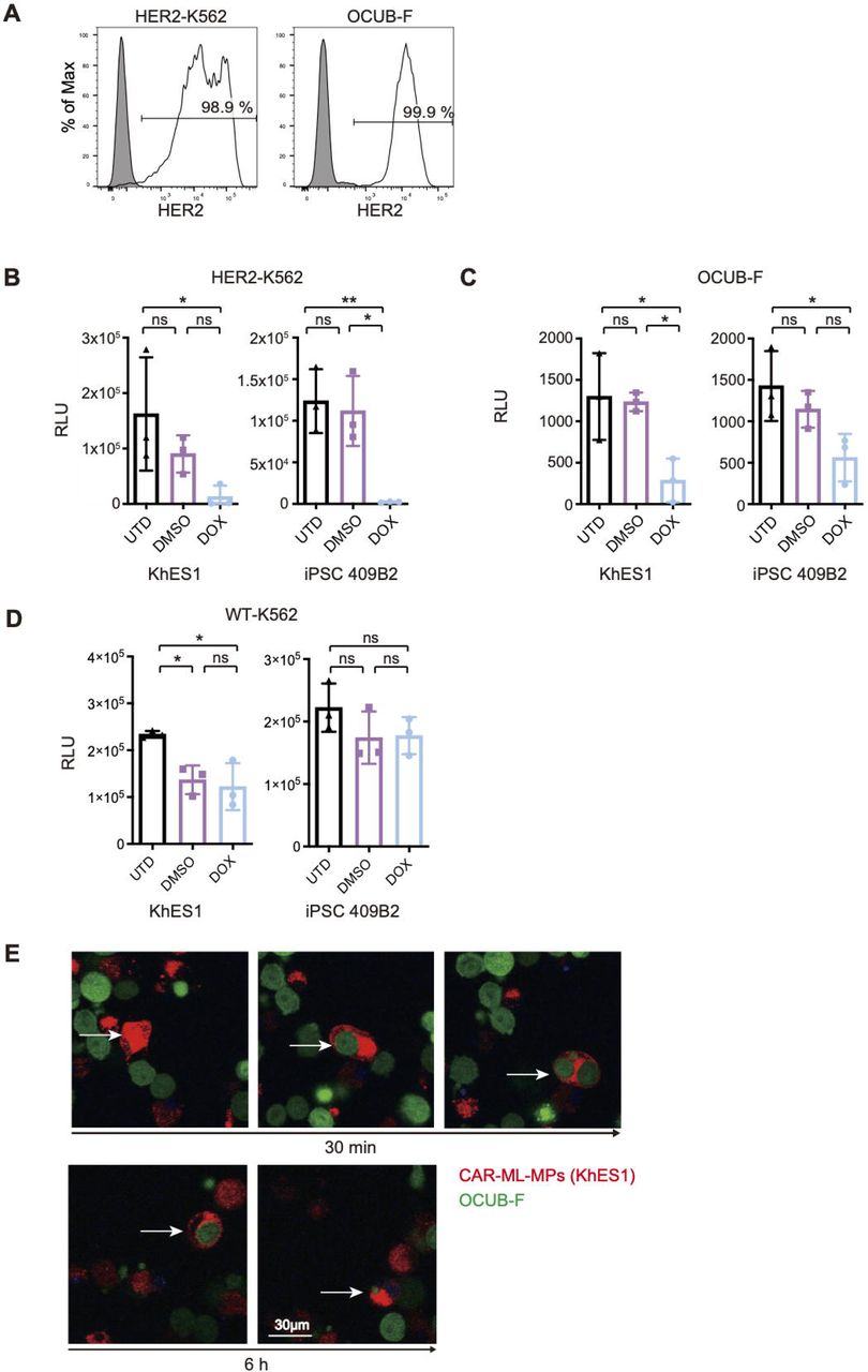 Human Her2 Erbb2 Protein, supplied by Sino Biological, used in various techniques. Bioz Stars score: 95/100, based on 1 PubMed citations. ZERO BIAS - scores, article reviews, protocol conditions and more https://www.bioz.com/product/biotinylated+mouse+anti+human+erbb2+her2/Human+HER2+%2F+ErbB2+Protein+(His+%26+AVI+Tag)%2C+Biotinylated/bio_rxiv__2024__07__22__604686-49-11-16 Average 95 stars, based on 1 article reviews
human her2 erbb2 protein - by Bioz Stars,
2026-11
95/100 stars
|
Buy from Supplier |
|
Eurofins
biotinylated pt-erbb2 peptide Biotinylated Pt Erbb2 Peptide, supplied by Eurofins, used in various techniques. Bioz Stars score: 90/100, based on 1 PubMed citations. ZERO BIAS - scores, article reviews, protocol conditions and more https://www.bioz.com/product/biotinylated+mouse+anti+human+erbb2+her2/u2os+erbb2+erbb4++osteosarcoma+cells/pmc04987620-107-11-22 Average 90 stars, based on 1 article reviews
biotinylated pt-erbb2 peptide - by Bioz Stars,
2026-11
90/100 stars
|
Buy from Supplier |
|
R&D Systems
biotinylated her2  Biotinylated Her2, supplied by R&D Systems, used in various techniques. Bioz Stars score: 95/100, based on 1 PubMed citations. ZERO BIAS - scores, article reviews, protocol conditions and more https://www.bioz.com/product/biotinylated+mouse+anti+human+erbb2+her2/Recombinant+Human+ErbB2%2FHer2+Fc+Chimera+Protein%2C+CF/pmc04550541-83-7-11 Average 95 stars, based on 1 article reviews
biotinylated her2 - by Bioz Stars,
2026-11
95/100 stars
|
Buy from Supplier |
|
Sino Biological
biotinylated mouse anti human erbb2 her2  Biotinylated Mouse Anti Human Erbb2 Her2, supplied by Sino Biological, used in various techniques. Bioz Stars score: 95/100, based on 1 PubMed citations. ZERO BIAS - scores, article reviews, protocol conditions and more https://www.bioz.com/product/biotinylated+mouse+anti+human+erbb2+her2/Anti-Human+Her2+ERBB2+Antibody%2C+Mouse+MAb/pmc07417204-42-1-29 Average 95 stars, based on 1 article reviews
biotinylated mouse anti human erbb2 her2 - by Bioz Stars,
2026-11
95/100 stars
|
Buy from Supplier |
Image Search Results
Journal: bioRxiv
Article Title: Treatment with anti-HER2 chimeric antigen receptor tumor-infiltrating lymphocytes (CAR-TILs) is safe and associated with antitumor efficacy in mice and companion dogs
doi: 10.1101/2022.09.11.507449
Figure Lengend Snippet: mRNA encoding anti-HER2 CAR was electroporated into TILs from five cutaneous melanomas (MM1, MM3, MM4, MM5, and MM6) and one uveal melanoma (UM22) to produce CAR-TILs. CAR expression was detected by HER2-biotin binding at 5-16 hours post-transfection (a). MM1 TILs and CAR-TILs were co-cultured with the parental or HER2 KO 92-1 uveal melanoma cell line for 4-6 hours, followed by flow cytometry to measure degranulation (CD107a expression) (b) or IFN-γ-secreting cells (c). TILs and CAR-TILs not co-cultured with 92-1 cells (TILs alone; TA) were used as controls in b-c. Alternatively, cells were co-cultured for 48 h, and the supernatant was collected for analysis of secreted IFN-γ by ELISA (d). 92-1 cells not co-cultured with TILs (no TILs) were used as a control. Data are presented as mean ± standard deviation (SD) of duplicates. The experiments were performed twice, and representative results are shown.
Article Snippet: For analysis of HER2 protein binding capacity of CAR-TILs, 100.000 cells were incubated with 1 μg
Techniques: Expressing, Binding Assay, Transfection, Cell Culture, Flow Cytometry, Enzyme-linked Immunosorbent Assay, Standard Deviation

Journal: bioRxiv
Article Title: Treatment with anti-HER2 chimeric antigen receptor tumor-infiltrating lymphocytes (CAR-TILs) is safe and associated with antitumor efficacy in mice and companion dogs
doi: 10.1101/2022.09.11.507449
Figure Lengend Snippet: Flow cytometry data showing HER2 CAR expression after HER2 CAR mRNA electroporation by detecting bound biotinylated HER2 protein. Representative plots for samples MM1, MM3, MM4, MM5, MM6 and UM22.
Article Snippet: For analysis of HER2 protein binding capacity of CAR-TILs, 100.000 cells were incubated with 1 μg
Techniques: Flow Cytometry, Expressing, Electroporation

Journal: bioRxiv
Article Title: Treatment with anti-HER2 chimeric antigen receptor tumor-infiltrating lymphocytes (CAR-TILs) is safe and associated with antitumor efficacy in mice and companion dogs
doi: 10.1101/2022.09.11.507449
Figure Lengend Snippet: (a-b) TILs and CAR-TILs from MM1 were co-cultured with the parental or HER2 KO 92-1 uveal melanoma cell line for 24 hours, followed by cleaved caspase 3 (CC3) detection in the tumor cells using flow cytometry (a). Additionally, TILs and CAR-TILs from MM5 were co-cultured with autologous cancer cells for 24 hours, followed by cleaved CC3 detection using flow cytometry (b). Cancer cells not treated with TILs were used as a control (no TILs). (c-g) Viability of autologous cancer cells was measured by luciferase signal detected after 48 hours co-culture with increasing doses of TILs and CAR-TILs from UM22 (c), MM3 (d), MM4 (e), MM5 (f) and MM6 (g) in the indicated ratios (TILs:cancer cells). Data is presented as mean with SD of triplicates. Asterisks represent p-values of difference between similar doses of TILs and CAR-TILs. The experiments were performed twice, and representative data from one experiment is shown.
Article Snippet: For analysis of HER2 protein binding capacity of CAR-TILs, 100.000 cells were incubated with 1 μg
Techniques: Cell Culture, Flow Cytometry, Luciferase, Co-Culture Assay

Journal: bioRxiv
Article Title: Treatment with anti-HER2 chimeric antigen receptor tumor-infiltrating lymphocytes (CAR-TILs) is safe and associated with antitumor efficacy in mice and companion dogs
doi: 10.1101/2022.09.11.507449
Figure Lengend Snippet: HER2 expression detected by qPCR (a) and immunohistochemistry (b) in tumors from four companion dogs (dogs 1-4). Canine cell lines D17.os and CF41.mg and canine PBMC were used as controls in a. (c) Expression of human melanoma marker Melan-a in tumors from dogs 3 and 4 with melanoma. (d) Classification of canine tumor biopsies from dogs 3 and 4 based on gene expression relative to TCGA cohort of > 10000 human tumor samples from 32 cancer types. The similarities between canine melanoma and samples in TCGA were visualized using tSNE dimensionality reduction. SKCM: melanoma cases. (e-f) TILs and mRNA electroporated CAR-TILs from dog 3 were co-cultured with the canine cell lines CF41.mg (e) and D17.os (f) for 24 h. The experiment was performed twice and representative data (n=3, data ± SD) from one experiment are shown. (g) Photograph of Dog 3 with metastatic oral malignant melanoma enrolled in the FIDO trial evaluating the safety of CAR-TIL treatment in companion dogs.
Article Snippet: For analysis of HER2 protein binding capacity of CAR-TILs, 100.000 cells were incubated with 1 μg
Techniques: Expressing, Immunohistochemistry, Marker, Cell Culture
Journal: mAbs
Article Title: Beyond bispecificity: Controlled Fab arm exchange for the generation of antibodies with multiple specificities
doi: 10.1080/19420862.2021.2018960
Figure Lengend Snippet: Generation of multispecific DB-VHH constructs. Trastuzumab IgG1-K409R mAb (anti-HER2) C-termini are fused to VHHs directed against EGFR, IL6R or NKG2D via a GS-linker. CS06 IgG1-F405L mAb (anti-c-MET) is fused to the same VHH molecules in the same manner. After recombinant production and purification, parental IgG-VHHs are recombined pairwise by reduction and reoxidation. The matching K409R and F405L mutations drive the generation of heterodimeric multispecific DB-VHHs.
Article Snippet: The biosensors were then rinsed in kinetics buffer (KB; PBS + 0.1% Tween-20 + 1% bovine serum albumin (BSA)) for 45 s (baseline) and the corresponding antigen was associated to the biosensor for 300 s. Different antigen concentrations (1:2 dilution rows in KB) were tested in order to identify the dynamic range of the binding kinetics and the most suitable concentration ranges were determined as follows:
Techniques: Construct, Recombinant, Purification

Journal: mAbs
Article Title: Beyond bispecificity: Controlled Fab arm exchange for the generation of antibodies with multiple specificities
doi: 10.1080/19420862.2021.2018960
Figure Lengend Snippet: Binding affinities measured for each paratope using recombinant antigens
Article Snippet: The biosensors were then rinsed in kinetics buffer (KB; PBS + 0.1% Tween-20 + 1% bovine serum albumin (BSA)) for 45 s (baseline) and the corresponding antigen was associated to the biosensor for 300 s. Different antigen concentrations (1:2 dilution rows in KB) were tested in order to identify the dynamic range of the binding kinetics and the most suitable concentration ranges were determined as follows:
Techniques: Binding Assay, Recombinant, Construct

Journal: mAbs
Article Title: Beyond bispecificity: Controlled Fab arm exchange for the generation of antibodies with multiple specificities
doi: 10.1080/19420862.2021.2018960
Figure Lengend Snippet: Biolayer interferometry analysis of simultaneous antigen binding of tri- and tetraspecific DB-VHHs. (a), (b) and (c) show exemplary sensorgrams for trispecific molecules and (d) a tetraspecific DB-VHH. The first association step represents binding of the DB-VHH (200 nM) via its CS06 paratope to biotinylated c-MET immobilized to streptavidin biosensors. Second (and third for (D)) association step is performed using an IL6R, EGFR or NKG2D recombinant protein (200 nM). The last association step is performed using HER2 (200 nM). Kinetic buffer (KB) controls were applied as negative controls for each association step.
Article Snippet: The biosensors were then rinsed in kinetics buffer (KB; PBS + 0.1% Tween-20 + 1% bovine serum albumin (BSA)) for 45 s (baseline) and the corresponding antigen was associated to the biosensor for 300 s. Different antigen concentrations (1:2 dilution rows in KB) were tested in order to identify the dynamic range of the binding kinetics and the most suitable concentration ranges were determined as follows:
Techniques: Binding Assay, Recombinant

Journal: mAbs
Article Title: Beyond bispecificity: Controlled Fab arm exchange for the generation of antibodies with multiple specificities
doi: 10.1080/19420862.2021.2018960
Figure Lengend Snippet: Specific cell clustering due to simultaneous binding of the DB-VHHs to three different cancer cell lines. Flow cytometry cytograms represent the fluorescence signals of the different cell populations. (a) Cells without antibody construct. Upper left gate = HCC-1954 (HER2 +++ ) cells stained with DeepRed, lower left gate = MDA-MB-468 (EGFR +++ ) cells stained with CMRA, lower right gate = EBC-1 (c-MET ++ ) cells stained with CFSE, upper right gate = HCC-1954 + EBC-1 cell doublets. Events in all three fluorescence channels (cell triplets) were marked in red. Cells were incubated in the presence of 1 nM (b) bispecific DB, (c) and (d) trispecific DB-VHHs, (e) and (f) tetraspecific DB-VHHs.
Article Snippet: The biosensors were then rinsed in kinetics buffer (KB; PBS + 0.1% Tween-20 + 1% bovine serum albumin (BSA)) for 45 s (baseline) and the corresponding antigen was associated to the biosensor for 300 s. Different antigen concentrations (1:2 dilution rows in KB) were tested in order to identify the dynamic range of the binding kinetics and the most suitable concentration ranges were determined as follows:
Techniques: Binding Assay, Flow Cytometry, Fluorescence, Construct, Staining, Incubation

Journal: mAbs
Article Title: Beyond bispecificity: Controlled Fab arm exchange for the generation of antibodies with multiple specificities
doi: 10.1080/19420862.2021.2018960
Figure Lengend Snippet: Simultaneous interaction of the DB-VHHs with HCC-1954 and EBC-1 target cells and binding of recombinant IL6R. Flow cytometry cytograms represent the fluorescence signals of the two cell populations and bound recombinant IL6R, detected with an anti-His6 detection antibody. (a) Cells without antibody construct. Upper left gate = HCC-1954 (HER2+++) cells stained with DeepRed, lower right gate = EBC-1 (c-MET++) cells stained with CFSE, upper right gate = HCC-1954 + EBC-1 cell doublets. Events in all three fluorescence channels (HCC-1954 + EBC-1 + bound recombinant IL6R-His Tag) were marked in blue. Cells were incubated in the presence of 10 nM (b) bispecific DB, (c) and (d) trispecific DB-VHHs, (e) and (f) tetraspecific DB-VHHs.
Article Snippet: The biosensors were then rinsed in kinetics buffer (KB; PBS + 0.1% Tween-20 + 1% bovine serum albumin (BSA)) for 45 s (baseline) and the corresponding antigen was associated to the biosensor for 300 s. Different antigen concentrations (1:2 dilution rows in KB) were tested in order to identify the dynamic range of the binding kinetics and the most suitable concentration ranges were determined as follows:
Techniques: Binding Assay, Recombinant, Flow Cytometry, Fluorescence, Construct, Staining, Incubation

Journal: mAbs
Article Title: Beyond bispecificity: Controlled Fab arm exchange for the generation of antibodies with multiple specificities
doi: 10.1080/19420862.2021.2018960
Figure Lengend Snippet: DB-VHHs elicit potent and specific NK cell-mediated target cell killing. Tumor cells were incubated with primary effector cells (NK cells) at a 1:5 ratio in the presence of antibody constructs in different concentrations. Error bars represent standard deviation of two biological replicates. Wildtype CS06 and trastuzumab with active Fc effector functioning were used as an ADCC reference (green). (a) and (b): c-MET-positive EBC-1 target cells, (c) and (d) HER2-overexpressing SK-BR-3 target cells. (a) and (c): NK cell cytotoxicity triggered by parental antibodies. (b) and (d): DB-VHHs serve as NK cell engager and mediate tumor cell killing.
Article Snippet: The biosensors were then rinsed in kinetics buffer (KB; PBS + 0.1% Tween-20 + 1% bovine serum albumin (BSA)) for 45 s (baseline) and the corresponding antigen was associated to the biosensor for 300 s. Different antigen concentrations (1:2 dilution rows in KB) were tested in order to identify the dynamic range of the binding kinetics and the most suitable concentration ranges were determined as follows:
Techniques: Incubation, Construct, Standard Deviation

Journal: mAbs
Article Title: Beyond bispecificity: Controlled Fab arm exchange for the generation of antibodies with multiple specificities
doi: 10.1080/19420862.2021.2018960
Figure Lengend Snippet:
Article Snippet: The biosensors were then rinsed in kinetics buffer (KB; PBS + 0.1% Tween-20 + 1% bovine serum albumin (BSA)) for 45 s (baseline) and the corresponding antigen was associated to the biosensor for 300 s. Different antigen concentrations (1:2 dilution rows in KB) were tested in order to identify the dynamic range of the binding kinetics and the most suitable concentration ranges were determined as follows:
Techniques: Variant Assay, Hydrophobic Interaction Chromatography, Mutagenesis, Molecular Weight, High Performance Liquid Chromatography
Journal: Pharmaceutics
Article Title: Biologic Evaluation of a Heterodimeric HER2-Albumin Targeted Affibody Molecule Produced by Chemo-Enzymatic Peptide Synthesis
doi: 10.3390/pharmaceutics14112519
Figure Lengend Snippet: ( A ) Overview of the Chemo-Enzymatic Peptide Synthesis (CEPS) method. Smaller peptide fragments are synthesized separately and subsequently joined by Omniligase. The detailed reaction scheme can be found in . ( B ) Schematic of PEP40233 produced by CEPS linking a variable fragment that is interchangeable for different binding specificities (here HER2) and a constant fragment comprising an ABD moiety for half-life extension. A chelating DOTA moiety may optionally be incorporated at the N-terminus during the chemical synthesis. ( C ) Schematic of the HER2 targeted Affibody molecule ABY-025 with a DOTA moiety conjugated at the C-terminus.
Article Snippet: The binding kinetics of
Techniques: Synthesized, Produced, Binding Assay

Journal: Pharmaceutics
Article Title: Biologic Evaluation of a Heterodimeric HER2-Albumin Targeted Affibody Molecule Produced by Chemo-Enzymatic Peptide Synthesis
doi: 10.3390/pharmaceutics14112519
Figure Lengend Snippet: ( A ) Single-cycle kinetics SPR sensorgrams showing PEP40233 concentration series (0.5, 1, 2, 4, 8 nM) interaction curve with, from left to right, immobilized Her2, HSA, and MSA. ( B ) A-B-A injection sensorgrams showing PEP40233 (8 nM) binding to HER2 in the presence of HSA/MSA. The data confirm binding to HER2 both in presence and absence of albumin.
Article Snippet: The binding kinetics of
Techniques: Concentration Assay, Injection, Binding Assay

Journal: Pharmaceutics
Article Title: Biologic Evaluation of a Heterodimeric HER2-Albumin Targeted Affibody Molecule Produced by Chemo-Enzymatic Peptide Synthesis
doi: 10.3390/pharmaceutics14112519
Figure Lengend Snippet: Kinetic parameters of PEP40233 interacting with HER2, HSA and MSA.
Article Snippet: The binding kinetics of
Techniques:

Journal: Pharmaceutics
Article Title: Biologic Evaluation of a Heterodimeric HER2-Albumin Targeted Affibody Molecule Produced by Chemo-Enzymatic Peptide Synthesis
doi: 10.3390/pharmaceutics14112519
Figure Lengend Snippet: In vitro specificity of [ 177 Lu]Lu-PEP40233 binding to ( A ) SKOV-3 and ( B ) BxPC3 cells in the presence and absence of HSA. Block refers to pre-saturation of HER2 receptors with unlabeled PEP40233. The data are presented as an average value from 3 samples ± SD. The data demonstrate specific binding to both cell lines, and both in the presence and absence of HSA.
Article Snippet: The binding kinetics of
Techniques: In Vitro, Binding Assay, Blocking Assay

Journal: Pharmaceutics
Article Title: Biologic Evaluation of a Heterodimeric HER2-Albumin Targeted Affibody Molecule Produced by Chemo-Enzymatic Peptide Synthesis
doi: 10.3390/pharmaceutics14112519
Figure Lengend Snippet: InteractionMap evaluation of the affinity of 177 Lu labeled PEP40233 binding to HER2-expressing SKOV-3 cells in the presence of HSA.
Article Snippet: The binding kinetics of
Techniques: Labeling, Binding Assay

Journal: Pharmaceutics
Article Title: Biologic Evaluation of a Heterodimeric HER2-Albumin Targeted Affibody Molecule Produced by Chemo-Enzymatic Peptide Synthesis
doi: 10.3390/pharmaceutics14112519
Figure Lengend Snippet: InteractionMap of [ 177 Lu]Lu-PEP40233 binding to HER2-expressing SKOV-3 cells in the presence of HSA. Binding was measured at concentrations of 0.25 nM, 0.75 nM, and 2.25 nM. Data are representatives from duplicates. The data show presence of two types of interactions with living HER2-expressing cells.
Article Snippet: The binding kinetics of
Techniques: Binding Assay, Expressing

Journal: Pharmaceutics
Article Title: Biologic Evaluation of a Heterodimeric HER2-Albumin Targeted Affibody Molecule Produced by Chemo-Enzymatic Peptide Synthesis
doi: 10.3390/pharmaceutics14112519
Figure Lengend Snippet: Biodistribution at 48 h post injection. ( A ) Biodistribution of [ 177 Lu]Lu-PEP40233 in BALB/C nu/nu mice bearing SKOV-3 and BxPC3 xenografts. Data are presented as an average value from four mice ± SD. The data show higher accumulation in xenografts with higher HER2 expression. ( B ) Comparison of biodistribution of [ 177 Lu]Lu-PEP40233 and [ 177 Lu]Lu-ABY-025 in SKOV-3 bearing xenografts. Data are presented as an average value from four mice ± SD. The data show clear reduction in the renal uptake and extension of residence in circulation for [ 177 Lu]Lu-PEP40233.
Article Snippet: The binding kinetics of
Techniques: Injection, Expressing, Comparison

Journal: Pharmaceutics
Article Title: Biologic Evaluation of a Heterodimeric HER2-Albumin Targeted Affibody Molecule Produced by Chemo-Enzymatic Peptide Synthesis
doi: 10.3390/pharmaceutics14112519
Figure Lengend Snippet: Imaging of mouse bearing SKOV-3 xenograft 48 h after injection of [ 177 Lu]Lu-PEP40233 using microSPECT/CT. Tumor (yellow arrow) and kidneys (white arrow) have elevated activity uptake, but tumor uptake exceeds the renal uptake. The scale is linear showing arbitrary units normalized to a maximum count rate.
Article Snippet: The binding kinetics of
Techniques: Imaging, Injection, Activity Assay
Journal: Nanomaterials
Article Title: The Precise Detection of HER-2 Expression in Breast Cancer Cell via Au 25 Probes
doi: 10.3390/nano12060923
Figure Lengend Snippet: Fluorescence imaging of BSA-biotin-fitc labeled HER-2 in SK cells by avidin and primary antibody-biotin at different time points.
Article Snippet: Human HER-2 antibody,
Techniques: Fluorescence, Imaging, Labeling, Avidin-Biotin Assay

Journal: Nanomaterials
Article Title: The Precise Detection of HER-2 Expression in Breast Cancer Cell via Au 25 Probes
doi: 10.3390/nano12060923
Figure Lengend Snippet: ( a ) Fluorescence imaging of 12 μM BSA-biotin-Au 25 labeled MDA-MB-231 cells at different time points. ( b ) HER-2 protein expressed in MDA-MB-231 cells before and after paclitaxel stimulation ( c ) Fluorescence imaging of BSA-biotin-Au 25 labeled MDA-MB-231 cells before and after paclitaxel stimulation.
Article Snippet: Human HER-2 antibody,
Techniques: Fluorescence, Imaging, Labeling
Journal: Advanced Pharmaceutical Bulletin
Article Title: A Systematic Study on Long-acting Nanobubbles: Current Advancement and Prospects on Theranostic Properties
doi: 10.34172/apb.2024.042
Figure Lengend Snippet: List of Nanobubble medications under clinical trials and in market
Article Snippet:
Techniques: Medications, Clinical Proteomics, Imaging, Over Expression
Journal: bioRxiv
Article Title: Enhanced anti-tumor effects through continuous administration of engineered CAR-macrophages derived from pluripotent stem cell-derived myeloid cell lines
doi: 10.1101/2024.07.22.604686
Figure Lengend Snippet: (A) Representative histograms showing HER2 expression in HER2-K562 and OCUB-F. (B, C) Cytotoxicity assay of CAR-ML-MPs derived from KhES1 and iPSC 409B2 strains against HER2-K562 (B) and OCUB-F (C). Effector-to-target ratio were 5:1 to HER2-K562 and 3:1 to OCUB-F, respectively (n = 3, independent experiment). UTD indicates untreated group. DMSO and DOX represent conditions treated without and with induction of CAR expression, respectively. RLU: relative light unit. Dunnett’s tests were performed as an a priori multiple comparison. *P < 0.05, **P < 0.01. Error bars represent mean ± standard deviation (SD). (D) Cytotoxicity assay of CAR-ML-MPs against wild type K562. (E) Results of time-lapse imaging of co-culture of CAR-ML-MPs (KhES1 strain) and OCUB-F (E/T ratio 1:1) after 30 minutes and 6 hours. Representative ML-MP cell (white arrow) showed phagocytosis of OCUB-F. See Video S1.
Article Snippet: For confirmation of anti-HER2 CAR expression, primary staining was performed with
Techniques: Expressing, Cytotoxicity Assay, Derivative Assay, Comparison, Standard Deviation, Imaging, Co-Culture Assay

Journal: bioRxiv
Article Title: Enhanced anti-tumor effects through continuous administration of engineered CAR-macrophages derived from pluripotent stem cell-derived myeloid cell lines
doi: 10.1101/2024.07.22.604686
Figure Lengend Snippet: (A) Experimental time course of in vivo assays comparing single and consecutive tumor treatments. (B) Bioluminescence images and (C) quantitative values of bioluminescence from Luc-HER2-K562 cells at the indicated days and conditions. Statistical analysis was performed using a priori Dunnett’s multiple comparisons test. UTD indicates untreated group; 1 and 1+6 indicate single treatment alone and with additional treatment, respectively. *P < 0.05, **P < 0.01. Error bars represent mean ± standard deviation (SD). (D) Kaplan–Meier plot showing the survival of each group (n = 3 for each group). Statistical analysis was performed with Log-rank (Mantel–Cox) test. (E) Experimental time course of repeated Luc-ML-MP transplantation assay. Bioluminescence images of (F) Luc-ML-MPs and (G) their quantification (n = 3). Error bars represent mean ± standard deviation (SD).
Article Snippet: For confirmation of anti-HER2 CAR expression, primary staining was performed with
Techniques: In Vivo, Standard Deviation, Transplantation Assay

Journal: bioRxiv
Article Title: Enhanced anti-tumor effects through continuous administration of engineered CAR-macrophages derived from pluripotent stem cell-derived myeloid cell lines
doi: 10.1101/2024.07.22.604686
Figure Lengend Snippet: (A) Schematic diagram of DOX-inducible anti-HER2 CAR construct. The construct includes HER2 recognition domain, intracellular hinge, CD28 and CD3ζ. TRE: tetracycline-response element. EF1: EF1 Alpha Promoter. TetR: Tet Repressor. ITR: inverted terminal repeat. (B) Proliferation curve of PSC-MLs derived from CAR-PSCs. On the left is mean ± standard deviation (SD) data for one passaging culture (n = 3, independent experiment). On the right, growth curves of KhES1-derived MLs cultured for 60 days. (C) Morphology of ML-MPs. Phase-contrast images (Upper panels) and May–Giemsa staining images (Lower panels). (D, E) Representative histograms showing macrophage cell-surface markers on CAR-ML-MPs. Panels (D) and (E) depict data from iPSC 409B2 and KhES1 strains, respectively. The rightmost panel displays histograms showing anti-HER2 CAR expression on the surface of CAR-ML-MPs in the presence of DOX. Gray fills represent isotype controls in all panels. (F) Expression of OX40L (left two panels) and 4-1BBL (right two panels). The relative quantity (Rq) of RNA expressions was measured in ML-MPs derived from the iPSC 409B2 strain (left) and the KhES1 strain (right) under both conditions, with and without DOX, compared to undifferentiated cells.
Article Snippet: For confirmation of anti-HER2 CAR expression, primary staining was performed with
Techniques: Construct, Derivative Assay, Standard Deviation, Passaging, Cell Culture, Staining, Expressing

Journal: bioRxiv
Article Title: Enhanced anti-tumor effects through continuous administration of engineered CAR-macrophages derived from pluripotent stem cell-derived myeloid cell lines
doi: 10.1101/2024.07.22.604686
Figure Lengend Snippet: (A) Experimental time course of in vivo tumor treatment assay. (B, C) Bioluminescence images (B) and quantitative values of bioluminescence (C) from Luc-HER2-K562 cells at the indicated days and conditions. Statistical analysis was performed using a priori Dunnett’s multiple comparisons test. UTD indicates untreated group. DMSO and DOX represent conditions treated without and with induction of CAR expression, respectively. *P < 0.05, ***P < 0.001. Error bars represent mean ± standard deviation (SD). (D) Kaplan–Meier plot showing the survival of each group (n = 5 for each group). Statistical analysis was performed with Log-rank (Mantel-Cox) test. (E) Experimental time course of Luc-ML-MP transplantation assay. Bioluminescence images of (F) Luc-ML-MPs and (G) their quantification (n = 3). Error bars represent mean ± standard deviation (SD).
Article Snippet: For confirmation of anti-HER2 CAR expression, primary staining was performed with
Techniques: In Vivo, Expressing, Standard Deviation, Transplantation Assay
Journal: Protein Engineering, Design and Selection
Article Title: Directed evolution of anti-HER2 DARPins by SNAP display reveals stability/function trade-offs in the selection process
doi: 10.1093/protein/gzv029
Figure Lengend Snippet: Primary screen of DARPins selected against HER2 using SNAP display. Four rounds of affinity selections were performed and 90 DARPins from the initial library, second and fourth round were tested for binding in a crude extract ELISA. Binding of DARPins to HER2 was detected using an HRP-conjugated antibody. The orange line indicates the ELISA signal of DARPin G3-HAVD in each screen. Over 90% of the clones screened gave a signal >5-fold over background while ∼30% of the selected DARPins showed greater binding signals compared with the parent DARPin G3-HAVD.
Article Snippet: The displayed protein library was incubated with
Techniques: Binding Assay, Enzyme-linked Immunosorbent Assay, Clone Assay

Journal: Protein Engineering, Design and Selection
Article Title: Directed evolution of anti-HER2 DARPins by SNAP display reveals stability/function trade-offs in the selection process
doi: 10.1093/protein/gzv029
Figure Lengend Snippet: ( A ) The frequency of mutations measured in the sequenced DARPin mutants is plotted onto the structure of DARPin H10-2-G3 (shown as putty cartoon): frequently mutated positions are colour-coded and rendered with backbone thickness proportional to mutation rate. Fully conserved positions are shown in dark blue. The position that accumulated ∼42% of sequence diversity, H52, is shown in red. The positions most frequently mutated are in proximity of the binding interface. Overall, positions mutated in greater than 10% of the sequenced population are all located in framework positions in the designed N2C DARPin library. Mutations do, however, map also on framework positions away from the binding interface. The interacting domain of HER2 is represented as a semi-transparent sand-coloured surface. ( B ) The published X-ray structure of DARPin H10-2-G3 (green) in complex with HER2 (blue) ( Jost et al. , 2013 ) was used to visualise the likely locations of the mutations obtained in DARPin F1. DARPin H10-2-G3 was affinity matured from the same parent clone (DARPin G3 HAVD) as the DARPins evolved by SNAP display. Mutations that contribute to binding are coloured brown while neutral mutations are coloured yellow (see Table ). Figures were prepared with Pymol based on PDB 4HRN ( Jost et al. , 2013 ).
Article Snippet: The displayed protein library was incubated with
Techniques: Mutagenesis, Sequencing, Binding Assay

Journal: Protein Engineering, Design and Selection
Article Title: Directed evolution of anti-HER2 DARPins by SNAP display reveals stability/function trade-offs in the selection process
doi: 10.1093/protein/gzv029
Figure Lengend Snippet: Affinities and binding kinetics of the selected HER2-binding DARPins a
Article Snippet: The displayed protein library was incubated with
Techniques: Binding Assay

Journal: Protein Engineering, Design and Selection
Article Title: Directed evolution of anti-HER2 DARPins by SNAP display reveals stability/function trade-offs in the selection process
doi: 10.1093/protein/gzv029
Figure Lengend Snippet: ( A ) ELISA analysis of selected DARPins. The top 10 DARPins (10 μg/ml of purified DARPin) were analysed for binding to immobilised HER2. All selected DARPins from SNAP display outputs gave a greater binding signal than DARPin G3-HAVD. All evolved DARPins were also analysed for their specificity in binding. No binding was observed to Streptavidin (which was used to capture biotinylated HER2 during selections) or lysozyme indicating the selected DARPins do not non-specifically bind to other proteins. Competition for binding of selected DARPins to HER2 with DARPin H10-2-G3 (10-fold excess DARPin H10-2-G3 over selected DARPins) showed that each selected DARPin competed for the same epitope as DARPin H10-2-G3. ( B ) Correlation between the observed fluorescence values of cell populations and the reciprocal of their binding affinity shows a correlation between the two properties. Most notably, Trastuzumab appears to have a significantly lower affinity on cells, probably as a result of the different binding mode and epitope the antibody binds to on HER2.
Article Snippet: The displayed protein library was incubated with
Techniques: Enzyme-linked Immunosorbent Assay, Purification, Binding Assay, Fluorescence
Journal: Analytical chemistry
Article Title: Rapid Enrichment and Detection of Extracellular Vesicles Enabled by CuS-Enclosed Microgels
doi: 10.1021/acs.analchem.9b04485
Figure Lengend Snippet: Quantification of EV concentrations with the 300-nm CuS-MG in 1× PBS or Serum. Calibration curves in 1×PBS using a) anti-CD63, b) anti-HER2; and in serum using c) anti-CD63 or d) anti-HER2. CL0 = chemiluminescence without EVs. [ABEI] = 0.5 mM, [H2O2] =1 mM, pH=11.
Article Snippet: The
Techniques:

Journal: Analytical chemistry
Article Title: Rapid Enrichment and Detection of Extracellular Vesicles Enabled by CuS-Enclosed Microgels
doi: 10.1021/acs.analchem.9b04485
Figure Lengend Snippet: Detection of cell released EVs using CuS-MG. The cell culture medium was collected from three cell lines: MCF-10A, MDA-MB-231 and SK-BR-3. a) Chemiluminescence resulted from EV detection in the culture media of three cell lines targeting CD63 and anti-HER2; b) PCA plot using the chemiluminescence data shown in a); c) Linear correlation between CD63 quantification results obtained by ELISA and the chemiluminescent signals from CuS-MG in our assay targeting CD63. [ABEI] = 0.5 mM, [H2O2] = 1 mM, pH=11.
Article Snippet: The
Techniques: Cell Culture, Enzyme-linked Immunosorbent Assay